Point-of-Care Ultrasound for Aesthetics & Plastic Surgery. Elevate Injection Safety and Surgical Precision with TodoPocus.
Which Ultrasound Scanner is Essential for Your Aesthetic and Reconstructive Procedures?
L20Pro
L20Pro Extended-Use Aesthetic Linear Ultrasound Probe for Skin & Injections
The Full-Face Navigator, Built for Full Clinic Days
- Probe Type: Linear Transducer
- Frequency: Linear: 16/20 MHz
- Scan Depth: Linear: 10-40mm
L20
L20 Aesthetic Linear Ultrasound Probe for Skin, MSK & Injection Guidance
The Facial Safety Standard
- Probe Type: Linear Transducer
- Frequency: Linear: 16/20 MHz
- Scan Depth: Linear: 10-40mm
L24Pro
L24Pro Extended-Use Facial Aesthetics Ultrasound Probe for Filler Mapping
The 24 MHz Microscope, Minus the Heat
- Probe Type: Linear Transducer
- Frequency: Linear 18/24 MHz
- Scan Depth: Linear: 10-25mm
L24
L24 Facial Aesthetics Linear Ultrasound Probe for Filler & Vessel Mapping
The 24MHz Microscope
- Probe Type: Linear Transducer
- Frequency: Linear 18/24 MHz
- Scan Depth: Linear: 10-25mm
L10H-Guide
L10H-Guide Linear Ultrasound Probe with Magnetic Navigation for Injections & Nerve Blocks
The Interventional Precision Tool
- Probe Type: Linear Transducer
- Frequency: Linear: 7.5/10 MHz
- Scan Depth: Linear: 20-100mm
D3-Ultra
D3-Ultra Premium 3-in-1 Handheld Ultrasound Scanner for Whole-Body POCUS
The Reconstructive Multi-Tool
- Probe Type: Linear, Convex, Phased Transducer 3-in-1
- Frequency: Convex: 3.2/5.0 MHz, Linear: 7.5/10 MHz, Phased: 3.2/5.0 MHz
- Scan Depth: Convex: 90-300mm, Linear: 20-100mm, Phased: 90-300mm
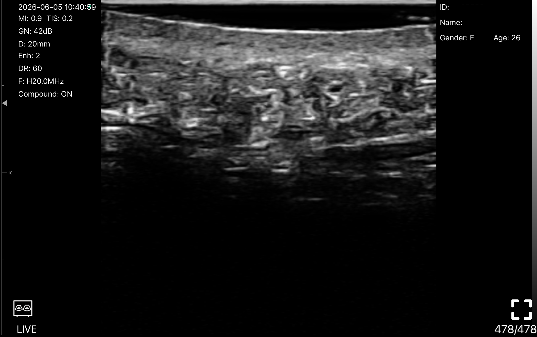
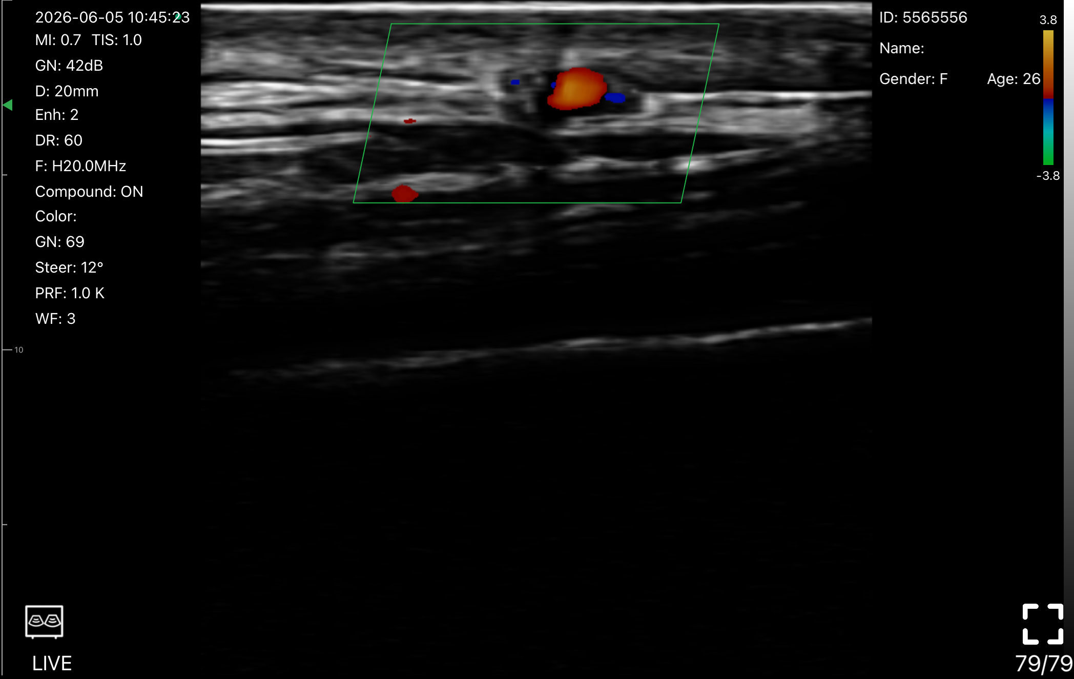
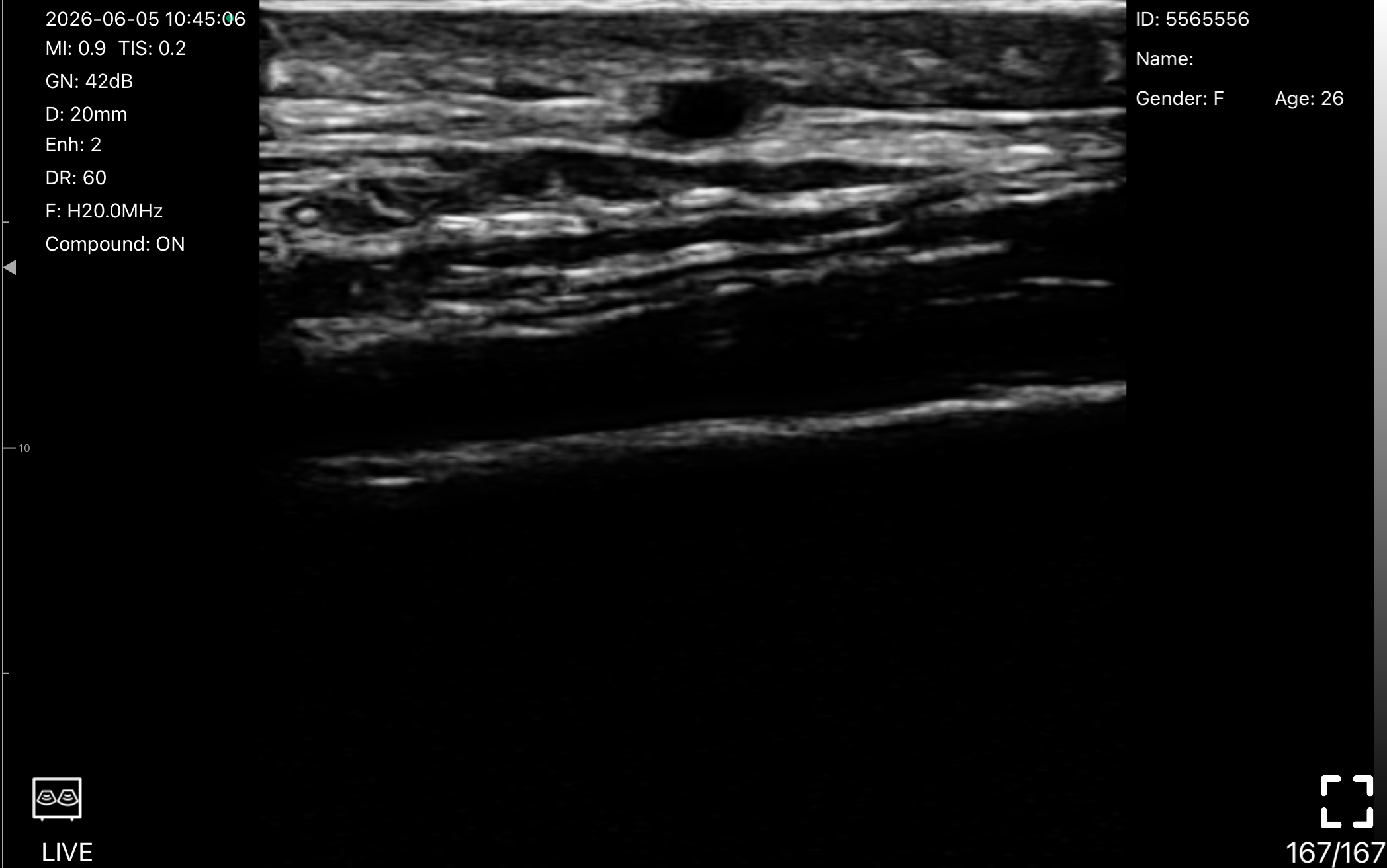
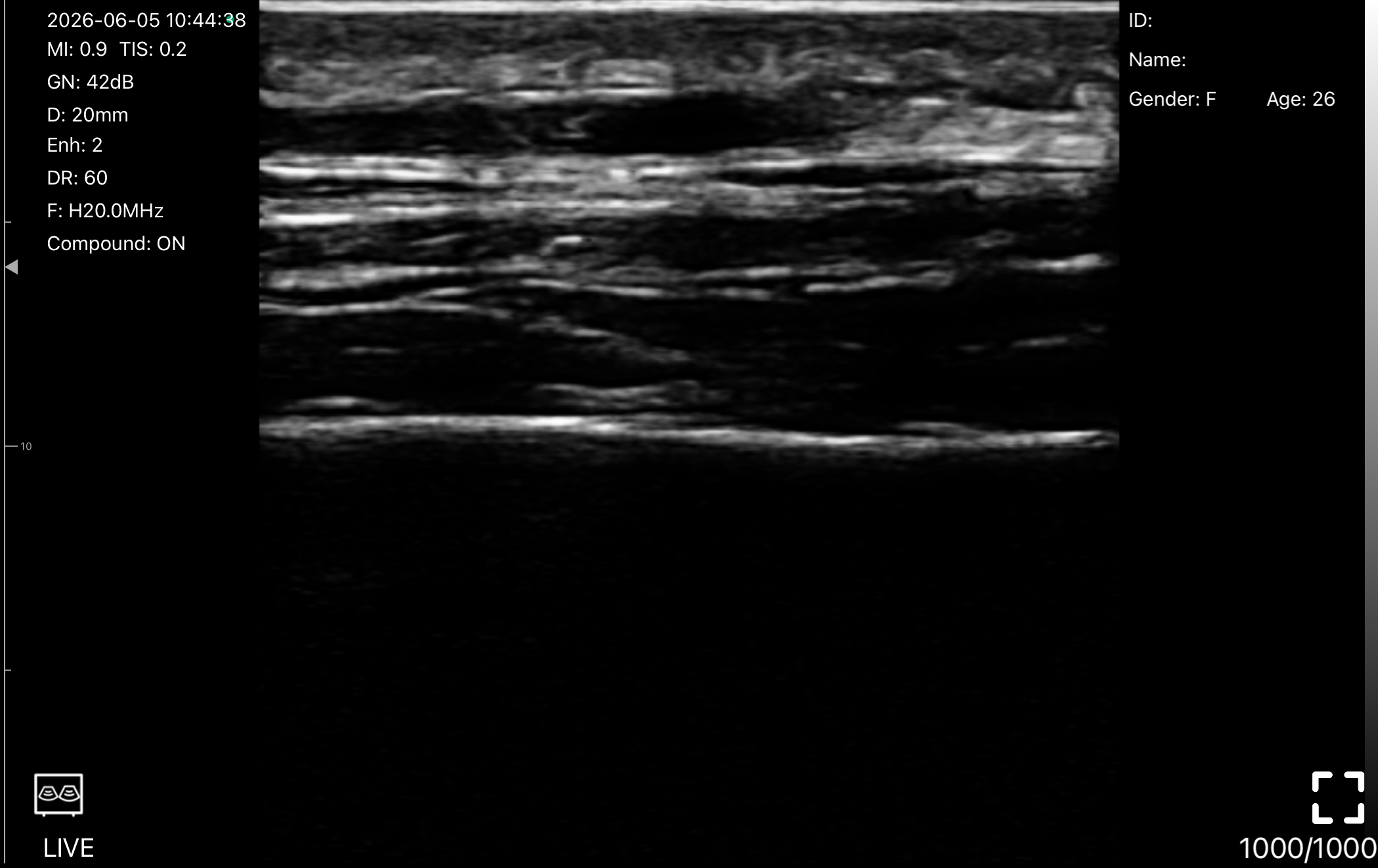
Ultra-High Frequency Mapping for Facial Aesthetics: Choose Depth, Detail, or Longer Scanning
For aesthetic clinicians, the right probe is not just about frequency. It is about seeing the anatomy that matters before every injection. TodoPocus L20/L20Pro and L24/L24Pro give you two focused paths: wider-depth facial mapping or ultra-superficial precision.
When your work moves across lips, cheeks, temples, jawline, and deeper soft-tissue planes, L20Pro gives you the range to keep up. Its 10-40mm scan depth helps you map vessels, locate filler, and understand anatomy across more treatment areas, while the Pro design adds longer battery life and improved thermal comfort for busy clinic days.
- Choose L20Pro when: You want the L20 imaging range with up to 3 hours of continuous scanning, a 4400mAh battery, and an optimized thermal shell for repeated aesthetic workflows.
- Choose L20 when: You want the same 16/20 MHz, 10-40mm imaging capability in a slimmer, lighter scanner for shorter sessions and daily portable use.
For delicate facial zones and ultra-superficial work, L24Pro brings your attention closer to the skin. Its 18/24 MHz imaging and focused 10-25mm range are built for fine vessel mapping, dermal and subcutaneous detail, and filler placement in shallow planes where every millimeter matters.
- Choose L24Pro when: You want surface-focused 18/24 MHz imaging with up to 3 hours of continuous scanning and a Pro shell built for longer, repeated use.
- Choose L24 when: You want the same 18/24 MHz, 10-25mm surface-detail capability in a lighter, slimmer scanner for flexible everyday use.
Clinical Evidence Gallery: See the Difference
Handheld Ultrasounds: Engineered for Global Standards
At TodoPocus, clinical safety and diagnostic reliability are our highest priorities. We strictly adhere to the most rigorous international medical regulations to ensure our technology supports you in every critical moment.
FDA 510(k) Cleared
CE Certified (Class IIa)
EU MDR 2017/745 Compliant
ISO 13485:2016
TGA Australia Approved
ANVISA Regulated
EUIPO Proprietary Innovation
HIPAA Compliant Security
One App. Three Platforms. Zero Subscription.
Turn your phone, iPad, Android tablet, or Windows PC into a premium diagnostic workstation. Download the app, connect wirelessly, and scan without limits.
Works on iOS, Android & Windows
Built-in Dedicated WiFi
DICOM & Storage Unlocked
5 Advanced Imaging Modes. Fully Unlocked.
Get standard access to premium features like Pulsed Wave (PW) Doppler and Color Flow. No subscriptions, no paywalls—just powerful diagnostics from day one.
B-Mode
High-Definition Grayscale. Visualize anatomical structures with superior contrast and edge detail.
M-Mode
Motion Analysis. Assess cardiac wall mechanics and fetal heart rates with high temporal resolution.
Color Doppler
Flow Dynamics. Rapidly identify blood flow direction and vessel patency in real-time.
Power Doppler (PDI)
High-Sensitivity Perfusion. Visualize low-velocity flow in microvasculature and hyperemic tissue.
Pulsed Wave (PW)
Spectral Quantification. Measure precise flow velocities to evaluate stenosis and vascular resistance.
17+ Specialized Presets. Instant Clinical Optimization. One-Tap Precision.
Streamline your exam with expert-tuned presets that instantly optimize frequency, depth, and gain for clinical precision—plus 3 fully customizable user slots to match your personal scanning preferences.
MSK
- Dynamic Exams: Real-time visualization of tendon gliding, ligament integrity, and muscle tears during joint movement.
- Inflammation: Power Doppler sensitivity to detect synovitis and active inflammation in joints.
- Injection Guidance: precise needle tracking for intra-articular injections and regenerative therapies.
Carotid
- Intima-Media Thickness: High-resolution borders to measure IMT and assess cardiovascular risk.
- Plaque Characterization: Identify calcified vs. soft plaque and screen for carotid stenosis.
- Stroke Prevention: Accurate velocity measurements to grade the severity of arterial narrowing.
Thyroid
- Nodule Characterization: High-frequency linear imaging to differentiate cystic vs. solid nodules and identify calcifications.
- Vascularity Assessment: Sensitive Color Doppler to evaluate hypervascularity ("Thyroid Inferno") in Graves' disease.
- Procedural Guidance: Crisp needle visualization for safe Fine Needle Aspiration (FNA) biopsies.
Vascular
- Deep Vein Thrombosis (DVT): Optimized for compression ultrasound to rule out clots in femoral and popliteal veins.
- Vessel Access: Clear visualization of vessel walls and compressibility for safe peripheral IV or PICC line placement.
- Flow Dynamics: Standard settings for assessing venous insufficiency and valve competency.
Skin
- Dermal Layers: Ultra-high frequency resolution to distinguish epidermis, dermis, and subcutaneous fat.
- Aesthetic Mapping: Visualize facial arteries and filler placement to prevent vascular occlusion during cosmetic procedures.
- Dermatology: Assess skin tumor depth, abscesses, or foreign bodies with sub-millimeter precision.
- Available on L20 & L24.
Small Parts
- Superficial Focus: Optimized focus zones for shallow structures like the testes, salivary glands, and lymph nodes.
- Acute Scrotum: Rapidly differentiate between testicular torsion (no flow) and epididymitis (hyperemia).
- Hernia Check: Dynamic imaging to visualize bowel loops within inguinal or ventral hernias during Valsalva.
Breast
- Mass Screening: Differentiate simple cysts from complex solid masses using high-definition grayscale contrast.
- Ductal Imaging: Visualize dilated ducts and intraductal pathologies.
- Bi-RADS Support: Clear margins and posterior acoustic features assist in accurate lesion classification.
Nerve
- Block Guidance: "Honeycomb" appearance enhancement to identify peripheral nerves for regional anesthesia (e.g., Brachial Plexus).
- Entrapment Scanning: Trace nerve paths to identify compression sites like Carpal Tunnel Syndrome.
- Safety: Clearly distinguish nerves from surrounding vessels to avoid accidental puncture.
Vessel Flow
- Low-Flow Sensitivity: Advanced Doppler algorithm designed to detect slow blood flow in small vessels or extremities.
- Perfusion Assessment: Evaluate tissue vascularization in diabetic foot ulcers or ischemic limbs.
- Hemodynamic Analysis: Clean spectral Doppler traces for precise velocity measurements in difficult-to-image vessels.
Abdomen
Deep Penetration: Penetrate up to 30cm to visualize deep abdominal organs (Liver, Kidney, Gallbladder) even in high-BMI patients.
Pathology Detection: Optimized contrast resolution for detecting hepatic steatosis, gallstones, and abdominal aortic aneurysms (AAA).
FAST Exam Ready: Rapidly assess free fluid in the hepatorenal (Morison’s) pouch and splenorenal recess during trauma triage.
Gynecology
- Pelvic Assessment: Visualize the uterus and adnexa with transabdominal clarity to assess for fibroids, cysts, or ectopic pregnancies.
- Fertility Monitoring: Clear visualization of ovarian follicles and endometrial lining thickness for IVF and fertility tracking.
- Acute Triage: Rapidly rule out ovarian torsion or ruptured cysts in emergency settings.
Obstetric
- Fetal Biometry: Optimized sound speed settings for accurate measurements of CRL, BPD, and FL to estimate gestational age.
- Wellbeing Assessment: Monitor fetal heart rate (FHR), amniotic fluid index (AFI), and placental position in real-time.
- Patient Connection: Crystal-clear 2D imaging to share the first look of the fetus with expectant parents.
Cardiac
- Real-Time Hemodynamics: High frame-rate imaging (Phased Array mode) to capture rapid valvular motion and wall contractility.
- Function Quantification: Supports rapid bedside assessment of Ejection Fraction (EF) and pericardial effusion.
- Critical Views: Optimized for clear Parasternal and Apical 4-Chamber views to rule out tamponade or gross dysfunction.
Urology
- Bladder Volume: Enhances fluid-to-tissue contrast for automated bladder volume calculation and residual urine assessment.
- Prostate Imaging: Visualize prostate size and shape to aid in BPH diagnosis.
- Urinary Tract: Quick scan of the ureterovesical junction (UVJ) to detect stones or jets.
Kidney
- Renal Architecture: Distinguish clearly between the renal cortex and collecting system to identify hydronephrosis.
- Stone Detection: High-contrast settings highlight hyperechoic calculi and their posterior shadowing.
- Perfusion Check: Sensitive Doppler settings to assess renal blood flow and rule out infarction.
Lung
- Artifact Analysis: Specifically tuned to highlight pleural lines, A-lines, and B-lines for pneumonia and pulmonary edema assessment.
- BLUE Protocol: Ideal for rapid bedside diagnosis of pneumothorax (Lung Point) vs. pleural effusion.
- COVID-19 & ARDS: Track lung consolidation and aeration changes in critical care patients.
Pediatrics
- Gentle Imaging: Reduced acoustic output power (ALARA principle) safe for pediatric and neonatal physiology.
- Fast & Clear: Motion-tolerant algorithms to capture diagnostic images quickly in moving children.
- Common Diagnoses: Optimized for ruling out pyloric stenosis, intussusception, and hip dysplasia.
What Are the Key Clinical Applications of Ultrasound in Plastic Surgery and Aesthetics?
Facial Aesthetics & Filler Complication Management
Reconstructive Surgery & Perforator Mapping
Breast Surgery & Body Contouring
I've been taking the D3-Ultra on my daily rounds, and it's genuinely useful. As you can see on the phone, the color flow is surprisingly sharp for a wireless handheld. No lag, no connection issues. Honestly, the best part is just keeping it in my coat pocket. It means I don't have to wait around for the hospital's main ultrasound cart, I just scan, get the answers I need, and move on.
I barely touch the ultrasound cart for quick checks now. The TodoPocus C5S lives in my pocket. Zero cables. I check AFI and fetal presentation right on my phone. The image is plenty clear to make the call.
Why Professionals Choose TodoPocus
The Freedom and Safety to Practice Medicine on Your Terms.
Freedom from Rent
Say goodbye to monthly fees. While other brands lock your device's core features behind expensive memberships, TodoPocus guarantees Lifetime Free software updates, DICOM features, and advanced Doppler modes. Our transparent pricing means your three-year total cost of ownership is over $2,000 less than leading competitors. Buy it once, own it forever.
| Device Model | TodoPocus D3-Ultra | Butterfly iQ3 | Clarius PAL HD3 | GE Vscan Air CL |
| Upfront Hardware | $3,470 | $3,899 | $5,395 | $4,999 |
| Probes Included | Linear + Convex + Phased | Single Probe for Whole Body | Phased + Linear | Convex + Linear |
| Annual Membership (Required for Adv. Features) | $0 (Included Forever) | $299 / year | $595 / year | Optional ($0) |
| 3-Year Total Cost | $3,470 | $4,796 | $7,180 | $4,999 |
| 5-Year Total Cost | $3,470 | $5,394 | $8,370 | $4,999 |
| You Save with TodoPocus | - | $1,924 | $4,900 | $1,529 |
The data is based on the official MSRP as of December 2025. Competitor subscription costs based on standard plans required for advanced Doppler/DICOM features.
The Math is Simple. Over 5 years, a "cheap" subscription probe can cost you double its sticker price. With TodoPocus, you pay $3,470 once. No monthly bills, no locked features, no surprises. Result: You save enough to buy a second TodoPocus for your backup bag.
Freedom from Cable
Cut the cord with zero-latency wireless connectivity and all-day battery life engineered for continuous scanning. Built to withstand the harshest clinical environments, our probes are IP67 waterproof, drop-tested, and ready for wherever the patient needs you.
Freedom from Platforms
Don't let your ultrasound probe dictate your hardware. TodoPocus is the only leading handheld solution offering native, plug-and-play support for iOS, Android and Windows system. Whether you are integrating into a hospital cart workflow or performing a quick bedside consult on your smartphone, our probe adapts to your ecosystem instantly—no specialized hardware required.
Uncompromising Privacy Secure
Patient data stays on YOUR device. We are HIPAA compliant by design because we never force your data onto third-party servers. Unlike competitors that mandate cloud uploads, TodoPocus empowers you with Local Storage. You own your data, you control where it goes, and you decide who sees it—ensuring total privacy and security.
Own Your POCUS with Zero Worry
World-class imaging technology, now within your reach.
30-Day Risk-Free Trial
Experience it in your clinic. Full refund if not satisfied.
3-10 Days Express Delivery
1-7 days to US, EU & AU. Delivery to your door globally within 3-10 days.
3-Year Warranty & Lifetime Support
Includes hardware repair plus lifetime free software & training.
Frequently Asked Questions about Point-of-Care Ultrasound for Aesthetics & Plastic Surgery
Choose by working depth. The L24 resolves the first 10–25 mm beneath the skin with 24 MHz detail — ideal if your work is strictly cutaneous: tear troughs, fine lines, skin-quality diagnostics, and ultra-superficial injections. The L20 trades a fraction of that surface resolution for a 40 mm working depth, letting you follow the facial artery at the piriform aperture, assess deep fat pads, and still define the SMAS clearly. If you buy one probe for full-face injectables, choose the L20 or L20Pro. If you never inject deeper than 2.5 cm, choose the L24 or L24Pro.
Imaging is identical — same transducers, frequencies, scan depths, and Doppler modes. The Pro Series upgrades endurance: a 4400 mAh battery doubles continuous scanning from 1.5 to 3 hours, and a heat-optimized Space Silver shell keeps the probe cool and comfortable through long vascular-mapping sessions, training days, and back-to-back clinics. If ultrasound is part of every consultation in your practice, the Pro pays for itself in uptime.
Standard software needle enhancement relies on acoustic reflection, which frequently fails during steep-angle insertions (>45°) or out-of-plane (OOP) approaches due to the physics of anisotropy. The L10H-Guide, however, utilizes passive magnetic sensors embedded in the probe to track a magnetized needle completely independent of the ultrasound beam. This projects a constant, real-time trajectory overlay on the screen, which clinical studies have shown reduces target distance errors by 57.1% and improves first-pass completion rates to 88.9% during complex nerve blocks.
Yes. TodoPocus wireless probes feature a minimum IP67 waterproof and dustproof design, with the transducer head specifically meeting IPX7 standards for liquid immersion. They are completely compatible with standard sterile surgical probe covers for use in the sterile field. Between cases, they can be safely and rapidly soaked or wiped down using hospital-approved high-level disinfectants and standard germicidal wipes, ensuring strict infection control compliance without risking internal electronic damage.
Ultra-high-frequency ultrasound reliably differentiates hyaluronic acid (HA) fillers, which appear as anechoic (dark) structures, from surrounding tissue and other biostimulators like calcium hydroxylapatite. This allows clinicians to perform precise, ultrasound-guided hyaluronidase injections directly into the HA deposit, rapidly restoring vascular perfusion and mitigating tissue necrosis safely.
Yes. High-frequency ultrasound readily distinguishes the sonographic signatures of different injectable products. Hyaluronic acid (HA) typically appears as well-defined, anechoic (black) fluid collections. In contrast, biostimulators like calcium hydroxylapatite (CaHA) or poly-L-lactic acid (PLLA) present as hyperechoic (bright) diffuse clouds, often with acoustic shadowing or bright particles. This diagnostic capability is invaluable when assessing unknown prior treatments or determining if a nodule can be effectively dissolved with hyaluronidase.
Aesthetic practitioners generally employ two complementary protocols: "Scan before injecting" and "Scan while injecting". The "Scan before" method is used prior to the procedure to map and mark the trajectory of critical vasculature, establishing safe injection zones. The "Scan while injecting" method utilizes real-time, in-plane imaging—often with a blunt cannula—to continuously visualize the instrument tip and the precise anatomical layer receiving the product, actively preventing intravascular placement.
Yes. Without ultrasound, clinicians must inject hyaluronidase blindly into the general area of suspected occlusion, often requiring multiple high-dose flooding treatments. Ultrasound guidance allows the practitioner to pinpoint the exact location and depth of the intravascular or compressive HA embolus. By targeting the hyaluronidase injection directly into the occlusion, clinicians can restore tissue perfusion much faster while utilizing significantly lower doses of the enzyme.
Unlock Clinical Clarity Anywhere.
Go wireless and subscription-free. Join 5,000+ clinicians
in the wireless POCUS revolution.
Unlock Clinical Clarity Anywhere.
Go wireless and subscription-free.
Join 5,000+ clinicians
in the wireless POCUS revolution.