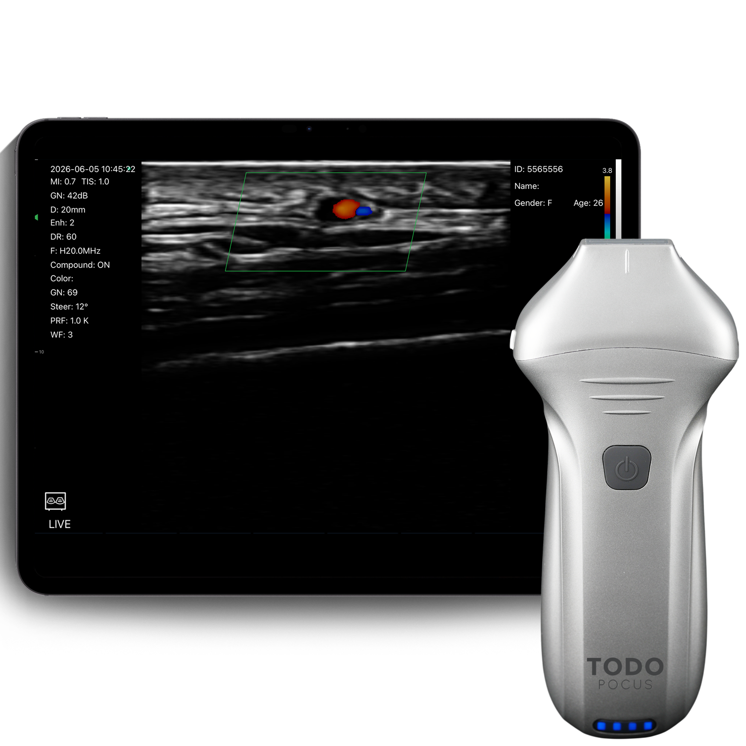
L20Pro Extended-Use Aesthetic Linear Ultrasound Probe for Skin & Injections
Estimated Arrival:
- US, EU, Oceania: 3-7 Business Days
- Rest of World: 5-10 Business Days
- All-Inclusive Shipping: We handle all customs clearance and cover taxes worldwide. Your order will be delivered directly to your door with no hidden fees or extra costs.
- Special Note for EU & South America: Please include your tax number (CPF/NIT/EORI) when placing your order. we'll ensure your package arrives safely, without any customs issues.
Enjoy worry-free delivery in TodoPocus with no customs delays or additional charges.
We process and ship all orders within 24 hours on business days. In the event of public holidays, shipping times may be slightly delayed. A notification will be sent to you in case of any changes to the shipping schedule.
Rest assured, we will prioritize your order and ensure it is shipped as soon as possible.
Your transactions are protected by SSL encryption, PCI-compliant payment gateways, and fraud detection systems. Check our Privacy Policy >
$5,990 Original price was: $5,990.$4,190Current price is: $4,190.
$0 Subscription Fees. Lifetime Free Updates.
Unlock the full diagnostic potential on iOS, Android, and Windows. No monthly bills, no cloud fees, and no feature lockouts—just unlimited scanning and seamless cross-platform compatibility forever.
TodoPocus L20Pro: 20 MHz Detail with Longer, Cooler Scanning.
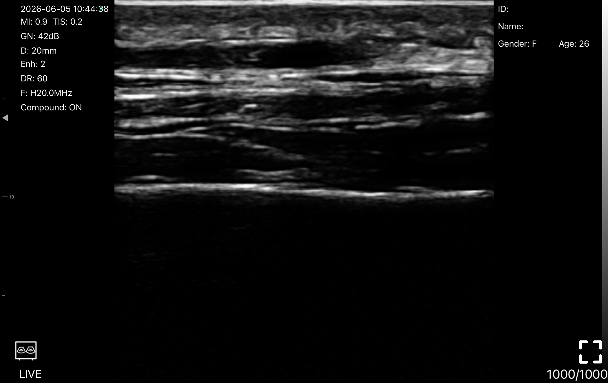
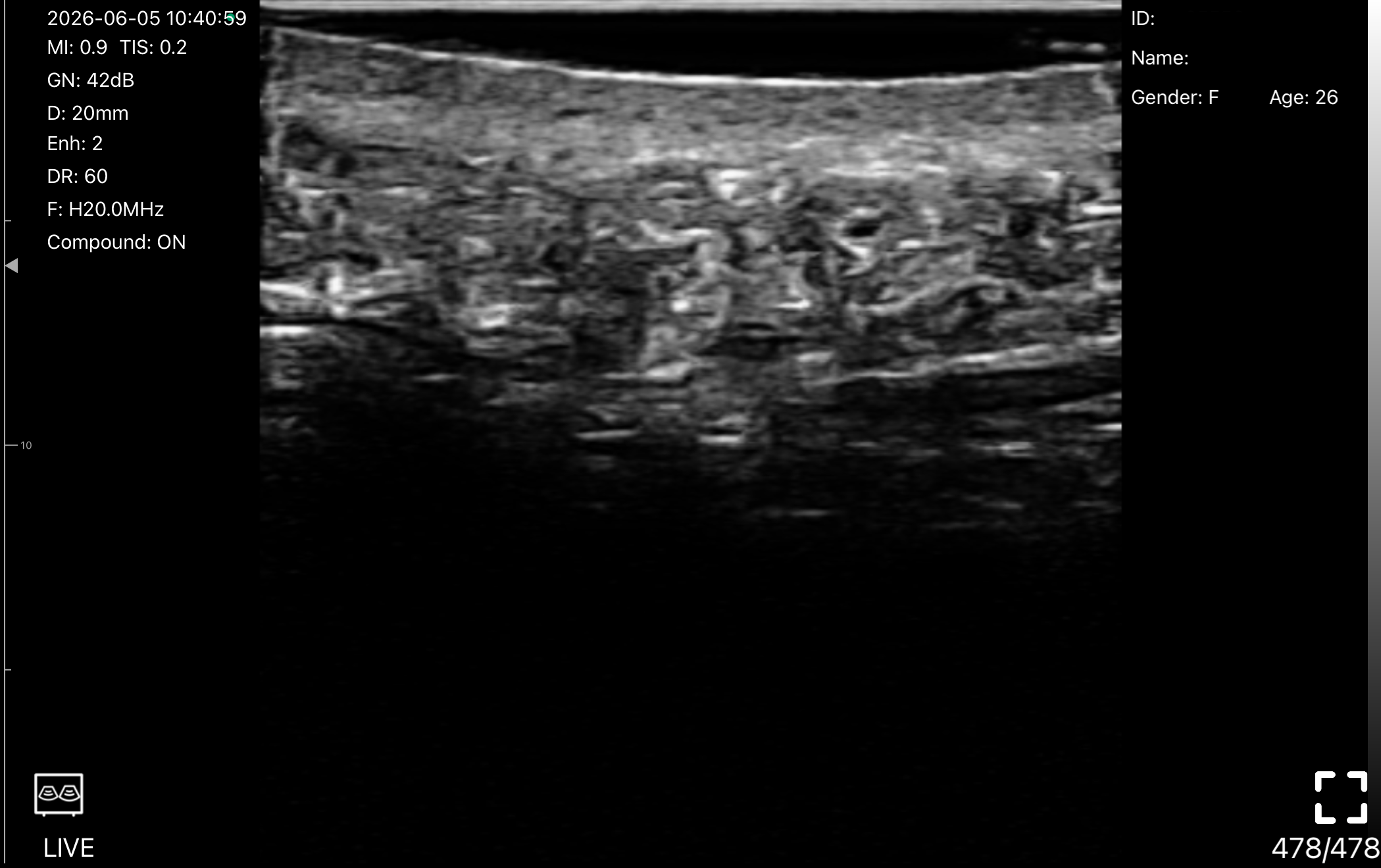
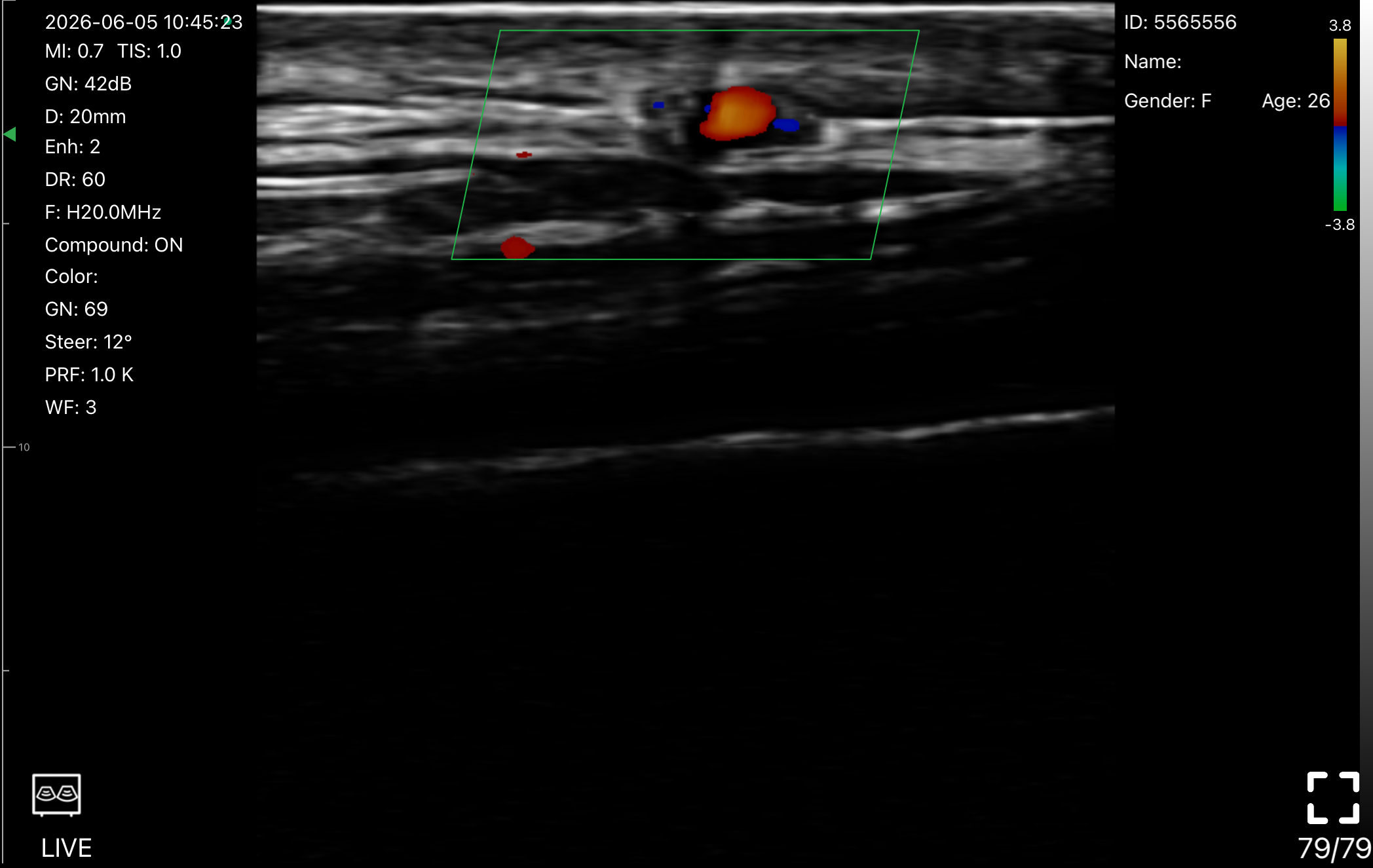
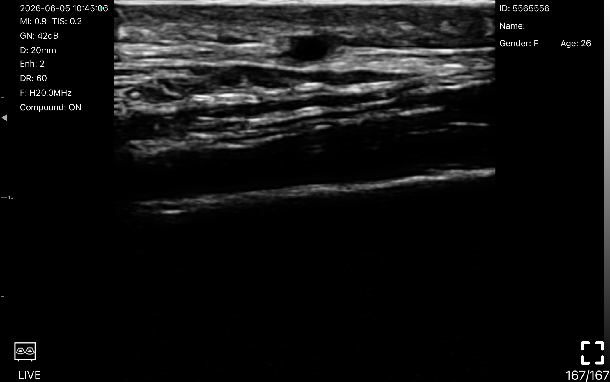
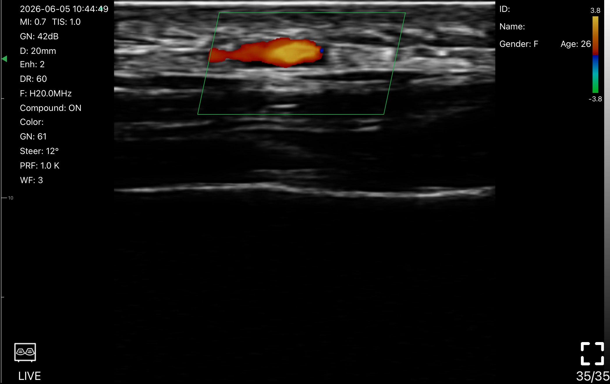
Visualize skin layers, facial vessels, filler placement, nerves, tendons, and superficial soft tissues with 16/20 MHz ultra-high-frequency linear imaging. Built with a 4400mAh battery and an optimized thermal Space Silver shell, L20Pro is designed for longer aesthetic, dermatology, MSK, and superficial vascular workflows with more stable scanning comfort.
Device Specifications: Uncompromising Performance. Pocket-Sized.
| Probe Type | Linear Transducer |
|---|---|
| Frequency | Linear: 16/20 MHz |
| Scan Depth | Linear: 10-40mm |
| Elements | 192 Elements |
| Channels | 64 Channels |
| Footprint | 25mm |
| Imaging Modes | B-Mode ,Color Doppler ,M-Mode ,Power Doppler ,Pulsed Wave Doppler |
| Shell | Optimized Thermal Space Silver Shell |
| Dimensions | 156mm x 60mm x 28mm |
| Weight | 218g |
| Battery | Built-in Lithium Battery, 4400mAh |
| Continuous Scanning Time | 3 Hours |
| Charging | USB Charging ,Wireless Charging |
| Cine Loop | 100, 200, 500, 1000 Frames |
| Connectivity | Built-in Wifi Wireless Connection ,Cable Connection |
| Mobile Platforms | iOS, Android, Windows |
| WiFi Type | 802.11n/2.4G/5G Dual-band 450 Mbps |
| Dynamic Range | 40/50/60/70/80/90/100/110 |
| Exported File Formats | JPEG, MP4, PNG, DICOM |
| Storage | Local Storage |
| Gain | 30-105 Db |
| Image Gray Scale | 256 Level |
| Noise Reduction | 0-1-2-3-4 |
| Puncture Assist | Flip ,Grid ,In-Plane Puncture Guide ,Out-of-Plane Puncture Guide |
| Doppler Imaging Adjustments | Color Doppler (Color, PDI): Steer, Color Gain (+/-), PRF (Pulse Repetition Frequency), Wall Filter (WF), Move & Zoom, Pulsed Wave Doppler (PW Mode): , PW Gain (+/-), Steer, Volume, Angle |
| General Imaging Adjustments | Depth, Dynamic Range (DYN), Enhancement (ENH), Focus, Frequency (F), Gain (GN), Harmonic Imaging, Noise Reduction, Time Gain Compensation (8TGC) |
| Software Measurements |
|
Handheld Ultrasounds: Engineered for Global Standards
At TodoPocus, clinical safety and diagnostic reliability are our highest priorities. We strictly adhere to the most rigorous international medical regulations to ensure our technology supports you in every critical moment.
FDA 510(k) Cleared
CE Certified (Class IIa)
EU MDR 2017/745 Compliant
ISO 13485:2016
TGA Australia Approved
ANVISA Regulated
EUIPO Proprietary Innovation
HIPAA Compliant Security
Clinical Evidence Gallery: See the Difference
Don't just take our word for it. Explore our library of high-resolution scans and clips demonstrating exceptional clarity across Cardiac, Abdominal, MSK, and Vascular applications.
One App. Three Platforms. Zero Subscription.
Turn your phone, iPad, Android tablet, or Windows PC into a premium diagnostic workstation. Download the app, connect wirelessly, and scan without limits.
Works on iOS, Android & Windows
Built-in Dedicated WiFi
DICOM & Storage Unlocked
5 Advanced Imaging Modes. Fully Unlocked.
Get standard access to premium features like Pulsed Wave (PW) Doppler and Color Flow. No subscriptions, no paywalls—just powerful diagnostics from day one.
B-Mode
High-Definition Grayscale. Visualize anatomical structures with superior contrast and edge detail.
M-Mode
Motion Analysis. Assess cardiac wall mechanics and fetal heart rates with high temporal resolution.
Color Doppler
Flow Dynamics. Rapidly identify blood flow direction and vessel patency in real-time.
Power Doppler (PDI)
High-Sensitivity Perfusion. Visualize low-velocity flow in microvasculature and hyperemic tissue.
Pulsed Wave (PW)
Spectral Quantification. Measure precise flow velocities to evaluate stenosis and vascular resistance.
17+ Specialized Presets. Instant Clinical Optimization. One-Tap Precision.
Streamline your exam with expert-tuned presets that instantly optimize frequency, depth, and gain for clinical precision—plus 3 fully customizable user slots to match your personal scanning preferences.
MSK
- Dynamic Exams: Real-time visualization of tendon gliding, ligament integrity, and muscle tears during joint movement.
- Inflammation: Power Doppler sensitivity to detect synovitis and active inflammation in joints.
- Injection Guidance: precise needle tracking for intra-articular injections and regenerative therapies.
Carotid
- Intima-Media Thickness: High-resolution borders to measure IMT and assess cardiovascular risk.
- Plaque Characterization: Identify calcified vs. soft plaque and screen for carotid stenosis.
- Stroke Prevention: Accurate velocity measurements to grade the severity of arterial narrowing.
Thyroid
- Nodule Characterization: High-frequency linear imaging to differentiate cystic vs. solid nodules and identify calcifications.
- Vascularity Assessment: Sensitive Color Doppler to evaluate hypervascularity ("Thyroid Inferno") in Graves' disease.
- Procedural Guidance: Crisp needle visualization for safe Fine Needle Aspiration (FNA) biopsies.
Vascular
- Deep Vein Thrombosis (DVT): Optimized for compression ultrasound to rule out clots in femoral and popliteal veins.
- Vessel Access: Clear visualization of vessel walls and compressibility for safe peripheral IV or PICC line placement.
- Flow Dynamics: Standard settings for assessing venous insufficiency and valve competency.
Skin
- Dermal Layers: Ultra-high frequency resolution to distinguish epidermis, dermis, and subcutaneous fat.
- Aesthetic Mapping: Visualize facial arteries and filler placement to prevent vascular occlusion during cosmetic procedures.
- Dermatology: Assess skin tumor depth, abscesses, or foreign bodies with sub-millimeter precision.
- Available on L20 & L24.
Small Parts
- Superficial Focus: Optimized focus zones for shallow structures like the testes, salivary glands, and lymph nodes.
- Acute Scrotum: Rapidly differentiate between testicular torsion (no flow) and epididymitis (hyperemia).
- Hernia Check: Dynamic imaging to visualize bowel loops within inguinal or ventral hernias during Valsalva.
Breast
- Mass Screening: Differentiate simple cysts from complex solid masses using high-definition grayscale contrast.
- Ductal Imaging: Visualize dilated ducts and intraductal pathologies.
- Bi-RADS Support: Clear margins and posterior acoustic features assist in accurate lesion classification.
Nerve
- Block Guidance: "Honeycomb" appearance enhancement to identify peripheral nerves for regional anesthesia (e.g., Brachial Plexus).
- Entrapment Scanning: Trace nerve paths to identify compression sites like Carpal Tunnel Syndrome.
- Safety: Clearly distinguish nerves from surrounding vessels to avoid accidental puncture.
Vessel Flow
- Low-Flow Sensitivity: Advanced Doppler algorithm designed to detect slow blood flow in small vessels or extremities.
- Perfusion Assessment: Evaluate tissue vascularization in diabetic foot ulcers or ischemic limbs.
- Hemodynamic Analysis: Clean spectral Doppler traces for precise velocity measurements in difficult-to-image vessels.
Abdomen
Deep Penetration: Penetrate up to 30cm to visualize deep abdominal organs (Liver, Kidney, Gallbladder) even in high-BMI patients.
Pathology Detection: Optimized contrast resolution for detecting hepatic steatosis, gallstones, and abdominal aortic aneurysms (AAA).
FAST Exam Ready: Rapidly assess free fluid in the hepatorenal (Morison’s) pouch and splenorenal recess during trauma triage.
Gynecology
- Pelvic Assessment: Visualize the uterus and adnexa with transabdominal clarity to assess for fibroids, cysts, or ectopic pregnancies.
- Fertility Monitoring: Clear visualization of ovarian follicles and endometrial lining thickness for IVF and fertility tracking.
- Acute Triage: Rapidly rule out ovarian torsion or ruptured cysts in emergency settings.
Obstetric
- Fetal Biometry: Optimized sound speed settings for accurate measurements of CRL, BPD, and FL to estimate gestational age.
- Wellbeing Assessment: Monitor fetal heart rate (FHR), amniotic fluid index (AFI), and placental position in real-time.
- Patient Connection: Crystal-clear 2D imaging to share the first look of the fetus with expectant parents.
Cardiac
- Real-Time Hemodynamics: High frame-rate imaging (Phased Array mode) to capture rapid valvular motion and wall contractility.
- Function Quantification: Supports rapid bedside assessment of Ejection Fraction (EF) and pericardial effusion.
- Critical Views: Optimized for clear Parasternal and Apical 4-Chamber views to rule out tamponade or gross dysfunction.
Urology
- Bladder Volume: Enhances fluid-to-tissue contrast for automated bladder volume calculation and residual urine assessment.
- Prostate Imaging: Visualize prostate size and shape to aid in BPH diagnosis.
- Urinary Tract: Quick scan of the ureterovesical junction (UVJ) to detect stones or jets.
Kidney
- Renal Architecture: Distinguish clearly between the renal cortex and collecting system to identify hydronephrosis.
- Stone Detection: High-contrast settings highlight hyperechoic calculi and their posterior shadowing.
- Perfusion Check: Sensitive Doppler settings to assess renal blood flow and rule out infarction.
Lung
- Artifact Analysis: Specifically tuned to highlight pleural lines, A-lines, and B-lines for pneumonia and pulmonary edema assessment.
- BLUE Protocol: Ideal for rapid bedside diagnosis of pneumothorax (Lung Point) vs. pleural effusion.
- COVID-19 & ARDS: Track lung consolidation and aeration changes in critical care patients.
Pediatrics
- Gentle Imaging: Reduced acoustic output power (ALARA principle) safe for pediatric and neonatal physiology.
- Fast & Clear: Motion-tolerant algorithms to capture diagnostic images quickly in moving children.
- Common Diagnoses: Optimized for ruling out pyloric stenosis, intussusception, and hip dysplasia.
27+ Clinical Measurements & Automated Tools: Built-in Standard.
From real-time hemodynamics to fetal biometry, access a complete suite of 27+ diagnostic calculation tools built directly into the app—no paid add-ons required.
Obstetrics & Gynecology (OB/GYN)
Comprehensive fetal biometry for confident prenatal assessment.
- Biometry Tools: Gestational Age via CRL, BPD, GS, FL, HC, AC.
- Calculations: Automated Estimated Fetal Weight (EFW) & Due Date (EDD).
- Fluid Assessment: Amniotic Fluid Index (AFI) for fetal well-being.
Cardiac & Hemodynamics
Advanced cardiac quantification at the bedside.
- Function: Ejection Fraction (EF) (via Teichholz/Simpson), Stroke Volume (SV), Cardiac Output (CO).
- Structure: Left Ventricular Internal Diameter (LVIDd/s) & Interventricular Septum.
- M-Mode: Precise Heart Rate (HR), Time, and Slope measurements.
Vascular & Flow Dynamics
Real-time hemodynamic analysis for vascular patency.
- Velocity Metrics: Peak Systolic (PS), End-Diastolic (ED), Time-Averaged Max (TAMAX), Time-Averaged Mean (TAMEAN).
- Indices: Resistive Index (RI), Pulsatility Index (PI), and S/D Ratio (Systolic/Diastolic).
- Volume Flow: Automatic Vessel Flow, Vessel Diameter, and Cross-Sectional Area.
General Imaging & Procedures
Versatile tools for everyday diagnostics.
- Core Tools: Distance, Trace (Manual Area), Ellipse (Area/Circumference), Angle.
- Urology: Automated Bladder Volume calculation (Post-Void Residual).
- Procedures: Needle Guide Trajectory (In-plane/Out-of-plane).
Split-Screen Compare: Validate Your Diagnosis in Real-Time
Stop toggling between saved images. Visualize the healthy anatomy alongside the pathology on a single screen. Whether you are comparing contralateral symmetry in MSK exams or tracking blood flow changes before and after an intervention, this mode gives you immediate visual confirmation without interrupting your scan.
One-Click Freeze: Capture the Perfect Image, Every Time
Instantly freeze the screen with a single click—either on the touchscreen or using the probe's physical button—to capture critical diagnostic images. This intuitive feature ensures you never miss a fleeting detail due to patient movement or shaky hands.
Needle Visualization Technology: Puncture with Absolute Confidence
Flexible File Exports with Notes: Save & Share in Seconds
Flexible Extra-Long Cine Loop: Review 1000 Frames of History
One Probe. Limitless Clinical Applications.
Select your specialty below to discover specific exams, presets, and workflows tailored to your practice.
- Aesthetics
- Dermatology
- Plastic Surgery
- Superficial MSK, Sports & Pain
- Superficial Anesthesia & Nerve Blocks
- Thyroid & Small Parts
- Superficial Breast & Chest Wall
- Vascular Access & Superficial Vessels
- Primary Care & Emergency
- Mobile Practice & Remote Consultation
- Research & Training
With 16/20 MHz high-frequency linear imaging and a 10–40 mm scan depth, the L20Pro helps aesthetic clinicians visualize facial vessels, filler placement, and superficial soft-tissue layers with clarity and Doppler flow insight.
- Map facial vessels before filler injections, correction procedures, and minimally invasive aesthetic treatments.
- Visualize filler location, filler migration, and tissue-plane relationships.
- Assess dermal, subcutaneous, and superficial fat layers for treatment planning.
- Review post-treatment changes such as swelling, nodules, or superficial filler-related complications.
- Use Color, Power, and PW Doppler to support safer injection planning and vascular-risk assessment.
The L20Pro supports dermatology workflows that require high-resolution imaging of skin, dermis, subcutaneous tissue, and superficial lesions within a focused 10–40 mm range.
- Evaluate superficial cysts, lipomas, abscesses, foreign bodies, and soft-tissue nodules.
- Measure lesion depth, margins, and relationship to surrounding superficial tissue.
- Assess dermal and subcutaneous thickness before treatment, biopsy, or excision planning.
- Use Doppler to evaluate vascularity around superficial inflammatory or suspicious areas.
- Monitor treatment response, wound healing, and post-procedural tissue changes over time.
Combining high-frequency detail, Doppler flow tools, and a 25 mm footprint, the L20Pro supports aesthetic and reconstructive workflows where superficial anatomy and tissue perfusion matter.
- Map superficial vessels before facial, scar, flap-adjacent, or skin-sparing procedures.
- Support filler correction, superficial fat grafting review, and soft-tissue reconstruction planning.
- Monitor postoperative seromas, hematomas, edema, or infection near incision sites.
- Evaluate superficial tissue perfusion patterns with Color, Power, and PW Doppler.
- Track scar maturation, graft-region changes, and healing response during follow-up.
With a 10–40 mm scan depth, MSK preset support, and Doppler sensitivity, the L20Pro is well suited for superficial musculoskeletal, sports medicine, physiotherapy, rheumatology, and pain-management workflows.
- Evaluate superficial tendons, ligaments, muscles, bursae, and small joints.
- Assess tendon gliding, ligament integrity, superficial muscle tears, and soft-tissue injury.
- Use Power Doppler to review hyperemia around inflamed tendons, joints, or entheses.
- Guide superficial injections, tendon sheath procedures, and small-joint interventions.
- Monitor healing after sports-related contusions, strains, and superficial soft-tissue injuries.
For anesthesia workflows, the L20Pro is best positioned for shallow peripheral nerve visualization, local anesthetic guidance, and superficial procedural support—not deep neuraxial or deep regional blocks.
- Visualize superficial nerves such as median, ulnar, radial, and other accessible peripheral nerve regions.
- Guide local anesthetic injections in shallow soft-tissue and minor procedural settings.
- Identify nearby vessels with Doppler before needle placement.
- Use grid, in-plane, and out-of-plane puncture guides for procedural confidence.
- Support superficial nerve block workflows where high-frequency detail and needle awareness are essential.
Compared with ultra-superficial aesthetic probes, the L20Pro’s 10–40 mm depth makes it better suited for selected thyroid, neck, and small-parts applications where high-frequency linear imaging is required.
- Evaluate superficial thyroid nodules, cystic structures, and gland architecture.
- Assess vascularity in superficial thyroid and neck-region findings with Color or Power Doppler.
- Support needle guidance for selected superficial thyroid or small-parts procedures.
- Examine superficial lymph nodes in the neck, axilla, or groin region.
- Document still images, cine loops, and DICOM files for follow-up or specialist review.
The L20Pro can support superficial breast and chest-wall imaging, especially for findings located close to the skin surface, postoperative review, and procedure-related follow-up.
- Evaluate superficial palpable findings within the 10–40 mm imaging range.
- Review peri-incisional seromas, hematomas, swelling, or superficial fluid collections.
- Assess superficial tissue changes after aesthetic or reconstructive breast procedures.
- Use Doppler to evaluate vascularity around shallow breast-region lesions.
- Save images and cine loops for documentation, follow-up, or consultation.
With Color, Power, and PW Doppler, plus dedicated vascular and vessel-flow presets, the L20Pro supports peripheral vascular access and selected superficial vascular assessment.
- Identify superficial peripheral veins before cannulation.
- Assess vessel depth, diameter, compressibility, wall structure, and surrounding tissue.
- Use Color or Power Doppler to confirm superficial blood flow and vessel patency.
- Support selected carotid, neck, and peripheral vessel workflows when anatomy falls within the 10–40 mm scan depth.
- Guide needle entry with grid, in-plane, and out-of-plane puncture support.
For frontline superficial POCUS, the L20Pro helps clinicians assess soft-tissue problems, minor injuries, and difficult vascular access without reaching beyond the limits of a high-frequency linear probe.
- Evaluate superficial abscesses, cellulitis, hematomas, and soft-tissue swelling.
- Localize superficial foreign bodies such as glass, splinters, or embedded debris.
- Assess superficial tendon, ligament, or soft-tissue injuries after minor trauma.
- Support ultrasound-guided peripheral IV placement in difficult-access patients.
- Capture and share images or cine clips for documentation, triage, and follow-up.
Wireless and cable connectivity, cross-platform compatibility, and extended battery capacity make the L20Pro practical for mobile superficial imaging and remote collaboration.
- Scan on iOS, Android, or Windows devices without subscription barriers.
- Capture high-resolution images and videos for case review or remote consultation.
- Export JPEG, PNG, MP4, and DICOM files for documentation and sharing.
- Support mobile aesthetics, dermatology, MSK, vascular access, and superficial soft-tissue workflows.
- Use the 4400 mAh battery and optimized thermal Space Silver shell for longer scanning sessions in clinic or mobile practice.
The L20Pro provides a flexible platform for training and research across superficial anatomy, Doppler imaging, procedural guidance, and high-frequency linear ultrasound workflows.
- Study superficial tissue layers, vessels, nerves, tendons, and small-parts anatomy within a 10–40 mm imaging range.
- Build training modules for ultrasound-guided aesthetics, dermatology, MSK, vascular access, and superficial procedures.
- Compare Doppler workflows for superficial vascularity, inflammation, and vessel-flow assessment.
- Document filler placement, soft-tissue changes, tendon movement, or procedural outcomes over time.
- Collect still images, cine loops, and DICOM files for education, protocol development, and longitudinal review.
Complete Scanner Kit: Everything You Need to Diagnose
- TodoPocus™ Handheld Ultrasound
- Premium Aluminum Carrying Case (Rugged & Lockable)
- Wireless Charging Pad
- USB-C & Lightning Cables
- Region-Specific Power Adapter
- Digital Quick Start Guide
Why Professionals Choose TodoPocus
The Freedom and Safety to Practice Medicine on Your Terms.
Freedom from Rent
Say goodbye to monthly fees. While other brands lock your device's core features behind expensive memberships, TodoPocus guarantees Lifetime Free software updates, DICOM features, and advanced Doppler modes. Our transparent pricing means your three-year total cost of ownership is over $2,000 less than leading competitors. Buy it once, own it forever.
| Device Model | TodoPocus D3-Ultra | Butterfly iQ3 | Clarius PAL HD3 | GE Vscan Air CL |
| Upfront Hardware | $3,470 | $3,899 | $5,395 | $4,999 |
| Probes Included | Linear + Convex + Phased | Single Probe for Whole Body | Phased + Linear | Convex + Linear |
| Annual Membership (Required for Adv. Features) | $0 (Included Forever) | $299 / year | $595 / year | Optional ($0) |
| 3-Year Total Cost | $3,470 | $4,796 | $7,180 | $4,999 |
| 5-Year Total Cost | $3,470 | $5,394 | $8,370 | $4,999 |
| You Save with TodoPocus | - | $1,924 | $4,900 | $1,529 |
The data is based on the official MSRP as of December 2025. Competitor subscription costs based on standard plans required for advanced Doppler/DICOM features.
The Math is Simple. Over 5 years, a "cheap" subscription probe can cost you double its sticker price. With TodoPocus, you pay $3,470 once. No monthly bills, no locked features, no surprises. Result: You save enough to buy a second TodoPocus for your backup bag.
Freedom from Cable
Cut the cord with zero-latency wireless connectivity and all-day battery life engineered for continuous scanning. Built to withstand the harshest clinical environments, our probes are IP67 waterproof, drop-tested, and ready for wherever the patient needs you.
Freedom from Platforms
Don't let your ultrasound probe dictate your hardware. TodoPocus is the only leading handheld solution offering native, plug-and-play support for iOS, Android and Windows system. Whether you are integrating into a hospital cart workflow or performing a quick bedside consult on your smartphone, our probe adapts to your ecosystem instantly—no specialized hardware required.
Uncompromising Privacy Secure
Patient data stays on YOUR device. We are HIPAA compliant by design because we never force your data onto third-party servers. Unlike competitors that mandate cloud uploads, TodoPocus empowers you with Local Storage. You own your data, you control where it goes, and you decide who sees it—ensuring total privacy and security.
Own Your POCUS with Zero Worry
World-class imaging technology, now within your reach.
30-Day Risk-Free Trial
Experience it in your clinic. Full refund if not satisfied.
3-10 Days Express Delivery
1-7 days to US, EU & AU. Delivery to your door globally within 3-10 days.
3-Year Warranty & Lifetime Support
Includes hardware repair plus lifetime free software & training.
Reviews: What the Medical Community is Saying
Sorry, no reviews match your current selections